Course
Study programs
Medical Studies in EnglishYear of study
2ISVU ID
ECTS
10
Histology and Embryology is a compulsory course at the second year of the Integrated Undergraduate and Graduate University Study of Medicine in English. It consists of 32 hours of lectures, 44 hours of seminars, and 44 hours of laboratory practicals, overall 120 hours (10 ECTS). Lectures are held in lecture halls of the Faculty of Medicine according to the course schedule. Seminars and laboratory practicals are held at the Department of Histology and Embryology.
Course objectives
Histology is one of the basic fields of medicine, which deals with the structure of a human body that can be studied using the light microscope or related devices. Histology also deals with the cell morphology (cytology) and with the fine structure of some organs (microscopic anatomy). Histology encompasses the entire microscopic and submicroscopic structure of an organism. Embryology studies the development of the embryo and helps students understand the complex relationship within the structure of the human body. Emphasis is on the morphogenesis during the organogenesis and on understanding the molecular and cellular basis of differentiation. Its practical medical implications are also of great importance since it accounts for the appearance of anomalies in the development of certain organs. Relationships between congenital malformations and errors in embryological development are discussed.
Expected course learning outcomes
At the end of this course, students will be able to demonstrate a working knowledge of human histology, development, correlate structure, and function of the human body. Students should be able to comprehend the molecular, biochemical, and cellular events that regulate the development of specialized cells, tissues, and organs during the embryonic development, tissue interactions and pattern formation, understand the experimental strategies and techniques that are used to identify the molecular and cellular mechanisms of development. Students should be thoroughly acquainted with structures and development of the human body by means of classical and contemporary methods of microscopic investigations and embryonic development; they should master the skills of microscopy of the most characteristic cells, tissues, and organs presented as histological slides. By utilizing their previous knowledge in physics, chemistry, biochemistry, biology, and anatomy, students should gain insight into the normal structure of the human body by means of light and electron microscopy.
Course content
The major role of histology in the medical curriculum is to provide basic understanding of many different aspects of structure and function of the human body. Emphasis is placed on the normal structure as a basis for proper functioning and for understanding pathophysiological processes. The following topics and subtopics will be considered: epithelial tissues (cellular membrane, basal lamina, cell-cell interactions); connective tissue (general characteristics, cells and intercellular substance, fibers, and ground substance); types of connective tissue (proper - dense, regular and irregular, adipose tissue); cartilage (hyaline elastic, fibrocartilage); bone (microscopic structure of bones, bone cells, histogenesis of bone, synovial membrane), blood, lymphocytes and their immune role; muscular tissue (smooth, skeletal, cardiac muscle), nervous tissue (structure of neuron, nerve fiber, synapse and the relationship of neurons, neuroglia, choroid plexus); blood vascular system, lymphatic system, endocrine system, respiratory 2 Assigned reading: Optional/additional reading: COURSE TEACHING PLAN: The list of lectures (with topics and descriptions): system, gastrointestinal tract, kidney and urinary tract, reproductive system, special senses.
The purpose of embryology is to provide students with a general outline of human development and to help them understand the complex relationship within the structure of the human body. Its practical medical implications are also of great importance since it can explain developmental anomalies and their molecular origins. The following topics and subtopic will be covered: fertilization, cleavage, gastrulation and formation of primary germ layers; differentiation of primary germ layers and organogenesis; cellular and molecular mechanisms that control tissue morphogenesis and differentiation; mechanisms that control differential gene expression leading to cell and tissue differentiation; extraembryonic coelom, connecting stalk, amnion, corium, placenta; neural plate, groove and tube; sex cycles, male and female sex organs; embryonic and fetal development; relationships between congenital malformations and errors in embryological development; environmental factors as causes of birth defects; development and anomalies of body systems; prenatal diagnostics.
1. A.L. Mescher.: Junqueira’s Basic Histology, XIV edition, The McGraw –Hill Education, New York 2016. 2. T.W.Sadler: Langman’s Medical Embryology, XIII edition, Wolters Kluwer Health, Philadelphia,2015. 3. http://medsci.indiana.edu/junqueira/virtual/junqueira.htm 4. https://accessmedicine.mhmedical.com/book.aspx?bookid=2430
Students are obliged to be prepared theoretically for seminars and practicals according to the executive education plan and this will be continuously checked. This course encourages discussion, individualized study, and work in small groups.
Class attendance, including test attendance, is mandatory. Students may be absent from 30% of each form of classes, provided they have a justifiable cause. If a student is absent for more than 30% of classes, they will have to re-enroll the course.
Students are expected to actively participate in all aspects of the course, complete reports from practicals on time, and attend the examinations. During LP, a student is obligated to have tools (a notebook, a blue and a red pencil, white coat).
As current epidemiological conditions do not allow direct teaching, it will be conducted according to the hybrid model as follows:
Lectures - recorded lectures that include individual teaching units will be available to the student on the Merlin platform of the course according to the schedule specified in the course syllabus. Seminars and laboratory practical - will take place in the lecture halls of the faculty according to the schedule in the syllabus with the use of histological images from the atlas and microscopic slides. In the case that students are unable to attend classes due to the deteriorating epidemiological situation, seminars and practical will be held in real time using the MS Teams platform following the group schedule listed in the syllabus.
Students will be able to come on-site for consultations before each partial test and before the final exam. Consultations will be held in the lecture halls of the faculty in the terms provided for lectures (in accordance with the epidemiological instructions). The schedule of consultations will be agreed with students. Consultations will also be organized on-site for the repetition of histological slides with the Institute's demonstrators. If necessary, all this consultation will be organized online.
Class attendance, including test attendance, is mandatory. Students may be absent from 30% of each form of teaching provided they have a justifiable cause. If a student is absent for more than 30% of the classes, they will have to re-enroll the course. Students are expected to actively participate in all aspects of the course, complete laboratory reports on time, and attend the examinations. Moreover, preparation of the course content, which is going to be discussed during seminars and laboratory practicals, is obligatory.
Student grading will be conducted according to the current Ordinance on Studies of the University of Rijeka (approved by the Senate) and the Ordinance on Student Grading at the Faculty of Medicine in Rijeka (approved by the Faculty Council).
Assessment of student work
Student work will be assessed and graded during the course and on the final exam. During the course, students may obtain a total of 100 grade points (credits). Students can achieve up to 70% of the final grade during the classes, and a maximum of 30% of the final grade at the final exam. Evaluation of students’ progress during classes, midterms, and the final exam in the academic year 2022/2023 is shown in Table 1.
Table 1. Distribution of grade points in the course “Histology and Embryology“
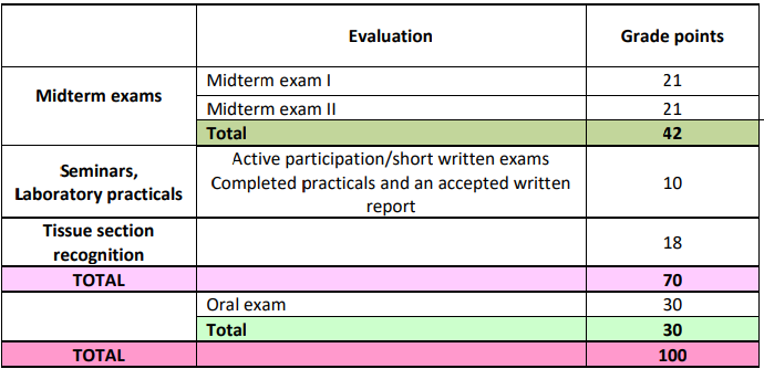
Written midterm exams
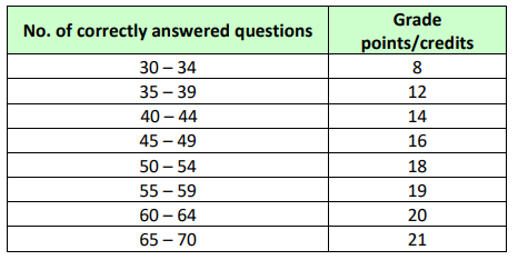
During the semester, two written midterm exams are planned that will include the content of lectures, seminars, and laboratory practicals. MT I – general histology and basic embryology. MT II – histology and embryology of various organs. 9 At each midterm exam, the maximum of grade points that a student can obtain is 21. All written midterm exams consist of 70 multiple-choice questions and are evaluated according to the criteria shown in Table 2.
MT I – during week 14. – 18.11.2022.
MT II – during week 23. – 27.01.2023.
Table 2. Evaluation of written midterm exams
Correction of the midterm exams
A student can access the correction of the midterm exams if they: i) did not obtain a minimum criteria (50% on each midterm) or ii) are not satisfied with the obtained credits and iii) in case of absence at the midterm exam due to a justified reason. If a student retakes the midterm exam because they are not satisfied with the obtained grade points, only the credits gained from the retaken midterms will be considered. Evaluation of the midterm corrections will be performed according to the criteria shown in Table 2. Students will have the opportunity to correct one or more midterm exam only once. Correction of the midterm exam I-II will be held after completing regular classes in terms set by the course schedule, before final exams in February and June.
Seminars and laboratory practicals
A student can obtain 10 credits (Table 3) throughout seminars and laboratory practicals after passing through 20 Topics (listed below). Evaluation of laboratory practicals implies a completed and accepted written report with all slides drawings. During laboratory practicals and seminars, the oral examination can be performed by the teacher or through short written exams.
Table 3. Evaluation of seminars and laboratory practicals
Topics T1 – Epithelial Tissue T2 – Connective Tissue, Blood, Cartilage, T3 – Bone, Osteogenesis, Bone Remodeling, Bone Marrow T4 – Muscle Tissue, Circulatory System T5 – Nerve Tissue, Nervous System T6 – Endocrine Glands T7 – Female Reproductive System, Gametogenesis, First Week of Development T8 – Second, Third Week of Development, Embryonic Period, Fetus, Fetal Membranes, Placenta, Twins T9 – Nervous System and Endocrine System Development T10 – Eye – Structure and Development T11 – Ear – Structure and Development 10 Other important information regarding to the course: T12 – Oral Cavity – Structure and Development, Teeth – Structure and Development T13 – Digestive Tract – Structure and Development T14 – Organ Associated with DT – Structure and Development T15 – Skin and Derivates – Structure and Development T16 – Immune System – Structure and Development T17 – Urinary System – Structure and Development T18 – Male Reproductive System – Structure and Development T19 – Respiratory System – Structure and Development T20 – Cardiovascular System – Development
Tissue section recognition
Is a compulsory oral exam and is required for students to be qualified for the final exam. A student must identify at least 10 of the 12 microscopic slides, as well as the structures that are described (and drawn) during the laboratory practicals, and can receive a maximum of 18 points. At least 9 points are required for passing the exam. Each slide is evaluated with ½, 1, or 1 ½ points depending on the student's knowledge. This exam will be held in the week before each final exam. At that time, the student can access the Tissue section recognition several times. Accurate dates and hours will be determined in agreement with the students.
Final exam
The final oral exam is mandatory and covers the entire course content. During the final exam, students can obtain a maximum of 30 credits. Assessment of the oral part of the final exam:
up to 15 credits: minimum criteria satisfied
16 – 20 credits: average criteria satisfied with noticeable errors
21 – 25 credits: answers with a few errors
26 – 30 credits: outstanding answers.
If a student is not satisfied with the final grade, they may refuse the grade. In case a student does not accept the grade, he/she must re-enter the final exam.
Conditions for admission to the final exam
A student who accomplishes 35 or more grade points during all course classes and/or after correction of the midterm exams, and passes Tissue section recognition with a minimum of 9 points can access the final exam.
A student who achieves less than 35 grade points during all course classes even after the correction of the midterm exams, or didn't achieve a minimum of 9 point on Tissue section recognition or is absent for more than 30% of all forms of classes, is graded as unsuccessful (F) and must re-enter the course.
Final grade
The final grade represents a sum of all grade points obtained during all course classes and on the final exam. Students are evaluated according to the ECTS (A-F) and numerical (5-1) system.
The ECTS and the numerical grading system are defined by the following criteria:
A (5) 90 – 100 credits
B (4) 75 – 89 credits
C (3) 60 – 74 credits
D (2) 50 – 59 credits
F (1) 0 – 49 credits
Class attendance, including test attendance, is mandatory. Students may be absent from 30% of each form of teaching provided they have a justifiable cause. If a student is absent for more than 30% of the classes, they will have to re-enroll the course. Students are expected to actively participate in all aspects of the course, complete laboratory reports on time, and attend the examinations. Moreover, preparation of the course content, which is going to be discussed during seminars and laboratory practicals, is obligatory.
Academic Honesty
It is expected that all students and teachers follow the Code of Academic Honesty in accordance with the Code of Ethics for the students of the Faculty of Medicine at the University of Rijeka. Please read the policy regarding academic honesty at: http://medical-studies-in-english.com/wp-content/uploads/2016/12/CODE-OF-ETHICS.pdf
Contact information
For questions and concerns, please feel free to contact us by e-mail or via the Department’s website. If you want to speak with a teacher during office hours (each working day between 11:00 am and 13:00 am), please let us know by e-mail or in class.
Expected competencies at course enrollment:
Students are expected to have basic knowledge of biology and anatomy.
Learning outcomes
To understanding the aim of the course. To recognize the role of Histology as a foundation for subsequent studies in pathology and physiology.
Learning outcomes
To define the microscopic structure and function of epithelial cells. To describe characteristic features of various types of epithelia.
Learning outcomes
To explain the types, characteristics, and functions of the connective tissue. To describe and to define cells and ground substance (fibers and basic substances) of connective tissue proper, and connective tissues with special properties. To define the peculiarities of microscopic and submicroscopic blood cells - erythrocytes, leukocytes, and platelets, and blood plasma. To adopt criteria for classification of blood cells based on their morphology.
Learning outcomes
To explain the classification, characteristics, and functions of supporting connective tissue. To define the ECM of different types of cartilage tissue. To explain the growth and healing processes of cartilage tissue damage. To explain the histological characteristics of joints. To explain the classification, characteristics, and functions of supporting connective tissue. To define the peculiarities of cells and bone matrix. To explain the characteristics of primary and secondary bone tissue with respect to their histological properties. To explain the processes of intramembranous and endochondral ossification. To describe features of fracture bone remodeling and repair.
Learning outcomes
To explain the classification, characteristics, and functions of three types of muscle tissue. To define cellular and ECM properties of smooth, skeletal, and cardiac muscle. To explain the ultrastructure of muscle fibers and morphological conditions for the possibility of contraction. To describe the histological structure of heart and vasculature.
Learning outcomes
To describe the classification, characteristics, and functions of the endocrine system. To define the specificity of the histological structure of certain endocrine glands; pituitary gland, epiphysis, thyroid, parathyroid glands, adrenal glands.
Learning outcomes
To explain the classification, characteristics, and functions of nerve cells (neurons and glial cells). To explain the processes of central and peripheral myelination. To define the cells and interstitial substances of certain parts of the central and peripheral nervous system (big and small brain, spinal cord, ganglia, peripheral nerves). To explain the ultrastructure of the nerve cells, the ability to transmit the signal, and the structure of the synapse. To describe the histological structure of meninges and blood-brain barrier.
Learning outcomes
To define the peculiarities of the histological structure of testes, epididymis and accessory glands. To describe the basis of key signaling pathways for development and some basis of organ formation. To understand and explain the processes of gametogenesis and the difference between spermatogenesis and oogenesis. |
Learning outcomes
To define the peculiarities of histological characteristics of the female reproductive system during different periods of a woman's life. To learn and adopt knowledge about sex cycles in males and females. To understand and explain changes during the generative period of life. To become familiar with the goal of learning developmental processes, fertilization, embryonic and fetal development of human embryos. To understand the underlying developmental processes: proliferation, migration, induction, differentiation and programmed morphogenic cell death To overcome the peculiarities of changes during the first week of development of the fertilized ovary (zygote). To outline the general changes during the second week (implantation, two-layered sham) and the third week (gastrulation) of development. To understand the main changes during the embryonic and fetal period of intrauterine development. |
Learning outcomes
To adopt knowledge about the development and function of fetal membranes: trophoblasts, amnions, coronas, egg yolks. To understand the development, texture, and function of the placenta and umbilicus in different periods of pregnancy. To understand the utero-placental bloodstream. |
Learning outcomes
To describe the structure and function of thin and thick skin layers. To understand and explain the structure of the skin glands and sensory receptors. To describe the main features of hair and nails. To explain diferent functional stages of the female mammary glands. To explain the developmental processes that allow the formation of individual skin layers and skin derivatives. |
Learning outcomes
To explain and describe the processes of the formation and differentiation of nerve and glial cells and the formation of nerve tissue during early neurogenesis. To understand the development of individual parts of the central and peripheral nervous system. To explain development of endocrine glands (pituitary, pineal, thyroid, parathyroid, adrenal). |
Learning outcomes
To define the histological structure of various parts of the external, middle, and internal ear. To understand the function of individual parts of the internal ear. To describe the developmental processes that enable the emergence of the outer, middle, and inner ear.
Learning outcomes
To define the peculiarities of the histological structure of the individual structures of the eye. To understand and explain the texture and function of the lens, cilia muscle, and individual parts of the retina. To explain the processes of optic cup development and formation of various parts of eye layers.
Learning outcomes
To explain the characteristics and functions of the immune system. To define the histological structure of the thymus, lymph nodes, spleen, and tonsils. To describe the developmental processes that lead to the formation of the lymph system organs. |
Learning outcomes
To define the general structure of the digestive tract. To describe the peculiarities of organs in the oral cavity (lip, tongue). To explain the structure of primary and permanent teeth. To explain the processes of denture formation in primary and secondary dentition. To define the processes that lead to tooth eruptions. |
Learning outcomes
To describe the peculiarities of organs - esophagus, stomach and intestine .
Learning outcomes
To define histological characteristics of parts forming the respiratory system (respiratory and olfactory region of the nose, paranasal sinuses, lungs, bronchi, bronchioles, alveoli). To understand and explain the structure and function of the blood-air barrier. |
Learning outcomes
To explain the basic characteristics of the structure and function of the urinary system. To define the peculiarities of the kidney structure - especially the cortex, the ureter, the bladder, the male and female urethra. To describe parts of the nephron. To define the characteristics of the transient epithelium. |
Learning outcomes
To understand the patterning of primary heart field, cardiac, and vascular development. To describe the developmental processes that lead to the formation of lymphatic capillaries and vessels. To describe the developmental processes of forming the respiratory system. |
Learning outcomes
To become familiar with the goal of learning developmental processes, fertilization, embryonic and fetal development of human embryos. To understand the underlying developmental processes: proliferation, migration, induction, differentiation and programmed morphogenic cell death |
To overcome the peculiarities of changes during the first week of development of the fertilized ovary (zygote). To outline the general changes during the second week (implantation, two-layered sham) and the third week (gastrulation) of development. To understand the main changes during the embryonic and fetal period of intrauterine development
Learning outcomes
To explain the basic facts of the development of histological techniques and microscopy. To get acquainted with and acquire knowledge about the way of preparing classic histological slides, as well as various histological, histochemical, and immunohistological techniques. To explain the principle of the methods used in histology laboratories and microscopy.
Learning outcomes
To classify and describe the microscopic and submicroscopic structure of epithelial cells. To define the peculiarities of certain types of glandular epithelia. (dental pulp - endothellium, small intestine – simple columnar, goblet cells, simple tubular glands, esophagus – squamous moist, mucous glands, skin – squamous dry, merocrine, holocrine, apocrine glands)
Learning outcomes
skeletal, cardiac, smooth muscle, endocardium, small artery and vein – HE, orcein staining
Learning outcomes
spinal cord and cerebellum – HE, silver staining, nerve, sensory, autonomic ganglia |
Learning outcomes
To learn about sex cycles in male and female sex. To understand and explain changes in histological structure in the ovaries and testes that precede the emergence of mature sex cells. (ovary, uterine tube, uterus, vagina)
Learning outcomes
thin skin with glands – axilla, hair, thick skin, mammary gland – 2 stages |
Learning outcomes
cornea, iris, ciliary body, lens, retina, development of eye – early, late stage |
Learning outcomes
lip, tongue, filiform and vallate papillae, dentin, cementum, enamel, enamel organ – early, late stage |
Learning outcomes
During seminars, students discuss in more detail themes introduced on the lectures and explain the unclear and insufficiently understandable topics. The seminars also provide an introduction to the topics that will be revealed on practicals. Students’ theoretical knowledge for each seminar is checked and students are therefore obliged to come prepared for this form of teaching. LP are followed by lecture topics or seminars that precede. The practical part of the practicals involves an overview of histological images of tissues and organs using microscopes and atlases mentioned in the literature list as well as drawing and a discussion with the teacher and demonstrator. The student is expected to be able to recognize the structures of various tissues and organs on microphotography, to be able to relate the observed details to the function of tissues or organs, and to be able to extract important characteristics of an unknown microscopic slide, compare with known structures and determine which organ or tissue is involved. 5 Students must have the appropriate drawing equipment (wooden pencils - red and blue) and a notebook (without lines) and white coats. Their participation in classes, understanding of the preparations, and their ability to recognize microscopic structures are evaluated in each LP. Thus, the student prepares to pass the Tissue Recognition Test at the end of the course, in which the same materials (atlas) will be used. In the description of learning outcomes for each seminar and LP, a list of histological slides is added.
To explain the basic facts of the development of histological techniques and microscopy. To get acquainted with and acquire knowledge about the way of preparing classic histological slides, as well as various histological, histochemical, and immunohistological techniques. To explain the principle of the methods used in histology laboratories and microscopy.
Learning outcomes
To describe histological characteristics of bone marrow. To understand the emergence of individual blood cells during intrauterine development, as well as the basis of the hematopoietic process later in life. |
Learning outcomes
To classify and describe the microscopic and submicroscopic structure of epithelial cells. To define the peculiarities of certain types of glandular epithelia. |
Learning outcomes
To explain the characteristics and functions of connective tissue. To define cells and ECM (fibers and ground substances), connective tissue, and connective tissues with special properties. To compare the similarities and differences between these two types of tissues.
|
Learning outcomes
To define the peculiarities of microscopic and submicroscopic structures of blood cells. To adopt criteria for blood cell definition based on their morphology. To define cellular and interstitial parts of different types of cartilage tissue. To explain the growth and healing processes of cartilage tissue damage. To explain the characteristics of the histological structure of joints. |
Learning outcomes
To define the peculiarities of cells and ECM of bone tissue. To explain the characteristics of primary and secondary bones with respect to their histological properties. To explain the processes of osteogenesis, the fracture healing process, and bone remodeling.
|
Learning outcomes
To define the peculiarities of stem cells and their cellular locations. To define the role of self-renewal and multipotency in stem cell biology. To explain the role of stem cells in the regeneration of tissues. To explain how differentiated tissues develop from stem cells. |
Learning outcomes
To clearly define cellular and interstitial properties of smooth, skeletal, and cardiac muscle tissue. To explain the ultrastructure of muscle cells and morphological conditions for the possibility of contraction in all types of muscle tissue. To describe the histological structure of the heart, artery, and vein. To adopt the classification of blood capillaries based on their microscopic structure. |
Learning outcomes
To explain the classification, characteristics, and functions of nerve cells (neurons and glial cells). To explain the processes of central and peripheral myelination. To define the cells and interstitial substances of certain parts of the central and peripheral nervous system (big and small brain, spinal cord, ganglia, peripheral nerves). To explain the ultrastructure of the nerve cells, the ability to transmit the signal, and the structure of the synapse. To describe the histological structure of meninges and the blood-brain barrier. |
Learning outcomes
To learn about sex cycles in male and female sex. To understand and explain changes in histological structure in the ovaries and testes that precede the emergence of mature sex cells. (ovary, uterine tube, uterus, vagina)
Learning outcomes
To explain and describe the processes that lead to the development of individual structures in the head and neck area. To adopt knowledge on the origin of the pharyngeal arches and its derivatives, the appearance of stomodeum and its differentiation during the embryonic and fetal developmental period. To describe the development of the temporomandibular joint.
Learning outcomes
To overcome the peculiarities of changes during the first week of development - zygote, pruning, second week – implantation, formation of a double layered shield. To overcome the peculiarities of changes during third week - embryonic, fetal development (neurulation, somitogenesis, germinal derivatives)
Learning outcomes
To explain the development of placental blood flow and function of embryonic envelopes – amnion, chorion, allantois, egg yolk sack. To understand the development, texture, and function of the placenta in different periods of pregnancy. |
Learning outcomes
To understand and explain the processes leading to differentiation of mesoderm and the formation of certain groups of skeletal and smooth muscles and the muscular wall of the heart. To explain the emergence of certain parts of the skeletal system - skull, spine, ribs, pelvis and limbs. |
Learning outcomes
To define the peculiarities of the histological structure and its development of testes, epididymis and accessory glands. |
Learning outcomes
To describe the classification, characteristics, and functions of the endocrine system. To define the specificity of the histological structure of certain endocrine glands; pituitary gland, epiphysis, thyroid, parathyroid glands and adrenal glands. |
Learning outcomes
To clearly define the peculiarities of the histological structure of the skin. To understand and explain the facts about the skin glands. To describe hair and nails. To explain developmental processes that allow the formation of individual skin layers and skin derivatives. To adopt knowledge about differences in appearance and function of the breast and breastfeeding between pregnant women, breastfeeding women and women that are not pregnant. |
Learning outcomes
To define the histological structure of various parts of the external, middle, and internal ear. To understand the function of individual parts of the internal ear. To describe the developmental processes that enable the emergence of the outer, middle, and inner ear. |
Learning outcomes
To understand and explain the structure and function of organs associated with the digestive tract – salivary glands, liver, pancreas and gallbladder. To understand and explain the flow of blood and bile inside the liver. |
Learning outcomes
To define the peculiarities of the histological structure of the individual structures of the eye. To understand and explain the texture and function of the lens, cilia muscle, and individual parts of the retina. To explain the processes of optic cup development and formation of various parts of eye layers. |
Learning outcomes
To explain the characteristics and functions of the immune system. To define the histological structure of the thymus, lymph nodes, spleen, and tonsils. To describe the developmental processes that lead to the formation of the organs associated with the lymphatic system. |
Learning outcomes
To understand the developmental processes of various organs of digestive tract formation |
Learning outcomes
To define the peculiarities of the individual parts of the oral cavity - lip, tongue, palate and teeth. To describe the development of the palate, the tongue, and the upper and the lower jaw. To explain the processes of denture formation in primary and secondary dentition.
Learning outcomes
To define the histological structure of certain parts of the digestive tract (esophagus, stomach, intestine, and colon). To understand and explain the structure and function of the individual layers in the structure of various segments. |
Learning outcomes
To understand and explain the structure and function of intestinal glands - salivary glands, liver, pancreas. To understand and explain the flow of blood and bile inside the liver. |
Learning outcomes
To define the basics of development and the peculiarities of the histological structure of the individual parts of the respiratory system (respiratory and nerve region, nose, paranasal sinuses, lungs, bronchi, bronchioles, alveoli). To understand and explain the structure and function of the blood-air barrier. (nasal cavity, trachea, lungs)
Learning outcomes
To explain the basic characteristics of the structure and function of the urinary system. To define the peculiarities of the kidney structure - especially the cortex, the ureter, the bladder, the male and female urethra. To describe parts of the nephron. To define the characteristics of the transient epithelium.
Learning outcomes
To understand and describe the processes that lead to the development of three generations of kidneys, the formation of the urethra, ureter, and urinary bladder. To understand the developmental process that leads to the normal male and female reproductive system - sexual glands and sex organs of the male and female sex. |
Learning outcomes
To define critical periods of development and to indicate teratogenic factors. To understand and explain the possibility of the emergence of anomalies and clinically important disorders that arise during development. |
| Academic year | |
|---|---|
| 2023/2024 | [Download] |